If treatment is indicated, diagnostic records to be evaluated during treatment planning are gathered. These include: facial and dental photographs, CT 3-dimensional x-ray, bite registration, and impressions of the teeth for study models. Dr. Suri evaluates each case and structures a treatment plan to achieve maximum result in minimal time.
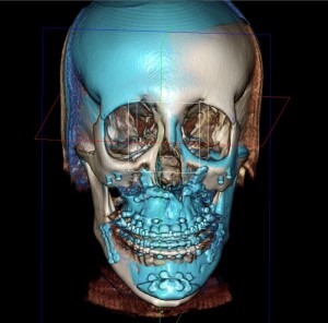
Register Two Scans for Superimposition Visualization on Orthognathic Cases
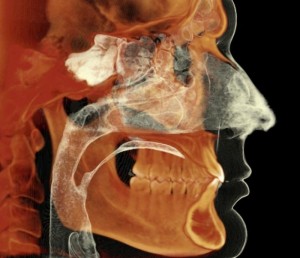
Capture Orthodontic Records in a Single Scan
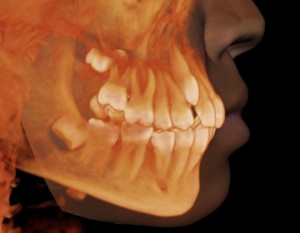
High Definition 3D Diagnostic Images for Ultimate Treatment Efficiency
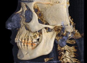
Capture Information Critical to Treatment and Control Radiation Exposure to the Patient
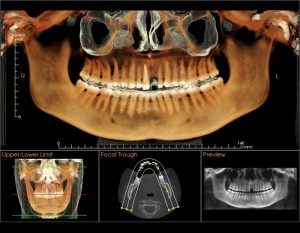
3D Panoramic Volume
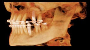
Low Dose Collimated Quick Scan
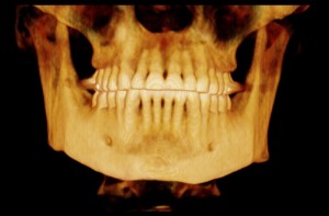
Low Dose Quick Scan
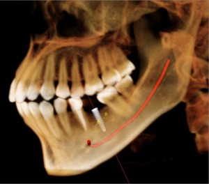
3D Views to Visualize Nerve Position for Implant Placement
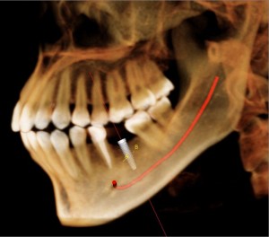
3D Views to Visualize Nerve Position for Implant Placement
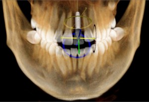
Plan Implant, Abutment and Final Restoration in One Software
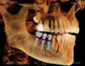
Restoration-Based Implant Planning
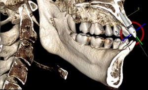
Visualize Implant within the Bone
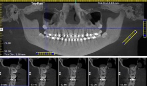
Views Help Identify Impacted Tooth Position, Relation to Other Teeth and Roots, as well as Pathology Prior to 3rd Molar Extractions
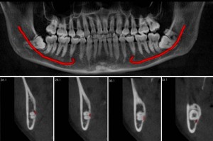
Clearly View the Relationship Between Impacted Tooth Roots and Nerve Prior to Extraction
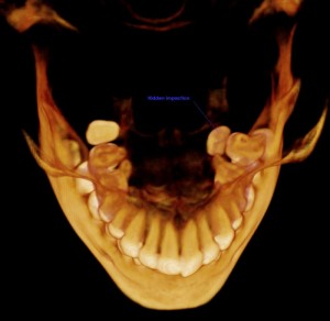
Reveal Hidden Impactions Not Seen on the Pan
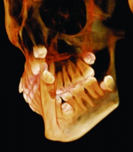
Supernumerary with a Full Crown
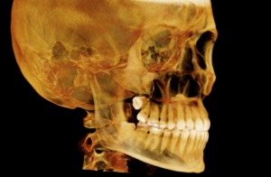
Extended Field of View
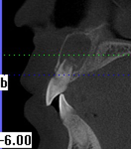
Identify Root Resorbtion in 3D that is Typically Undetected in 2D
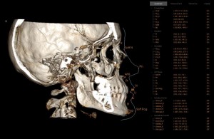
3D Cephalometric Analysis
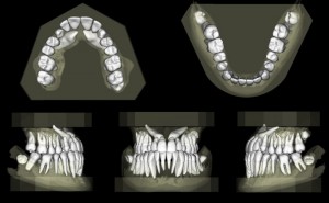
Digital Modeling
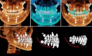
Orthodontic Treatment Simulation

Corrected Angle Views of Temporomandibular Joints
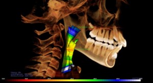
Airway Analysis
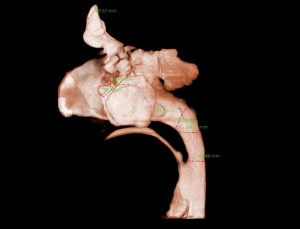
Sinus Evaluation

